Liquid-based cytology has revolutionized the way we examine cellular specimens, offering enhanced clarity and improved diagnostic capabilities. However, as with any diagnostic tool, attention to detail is paramount to accurate interpretation. In a recent case involving a 30-year-old woman, this principle proved crucial.
Upon initial examination, Figures 1 and 2 revealed cellular atypia suggestive of squamous intraepithelial lesion (LSIL). Key features such as karyomegaly and the presence of halos were evident, prompting concern for abnormal cellular proliferation. However, a deeper investigation uncovered a surprising twist.
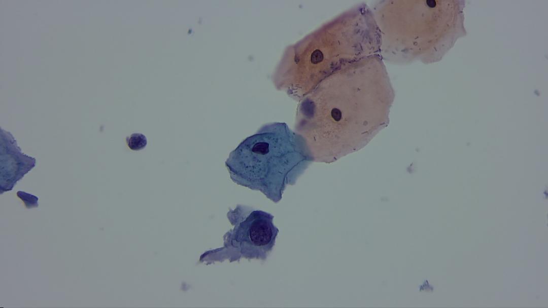
Fig. 1
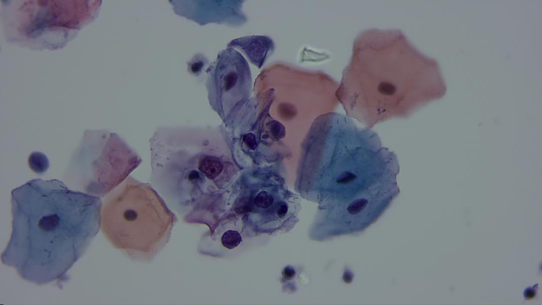
Fig. 2
Figures 3 through 8 unveiled a critical aspect overlooked in the initial assessment—trichomonas. These elusive organisms, known for their distinctive morphology, often masquerade as cellular abnormalities, leading to potential misdiagnosis. In this case, their presence obscured clear diagnostic clues, creating a diagnostic dilemma.
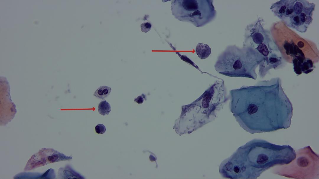
Fig. 3
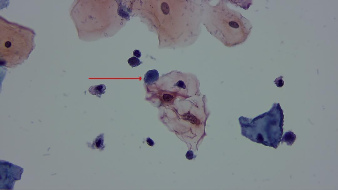
Fig. 4
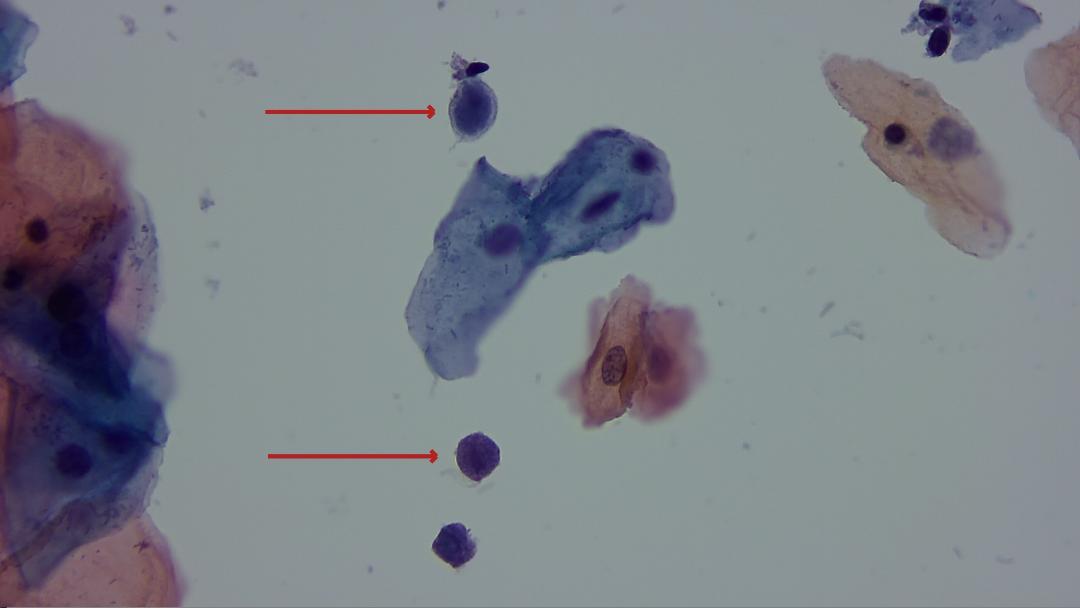
Fig. 5
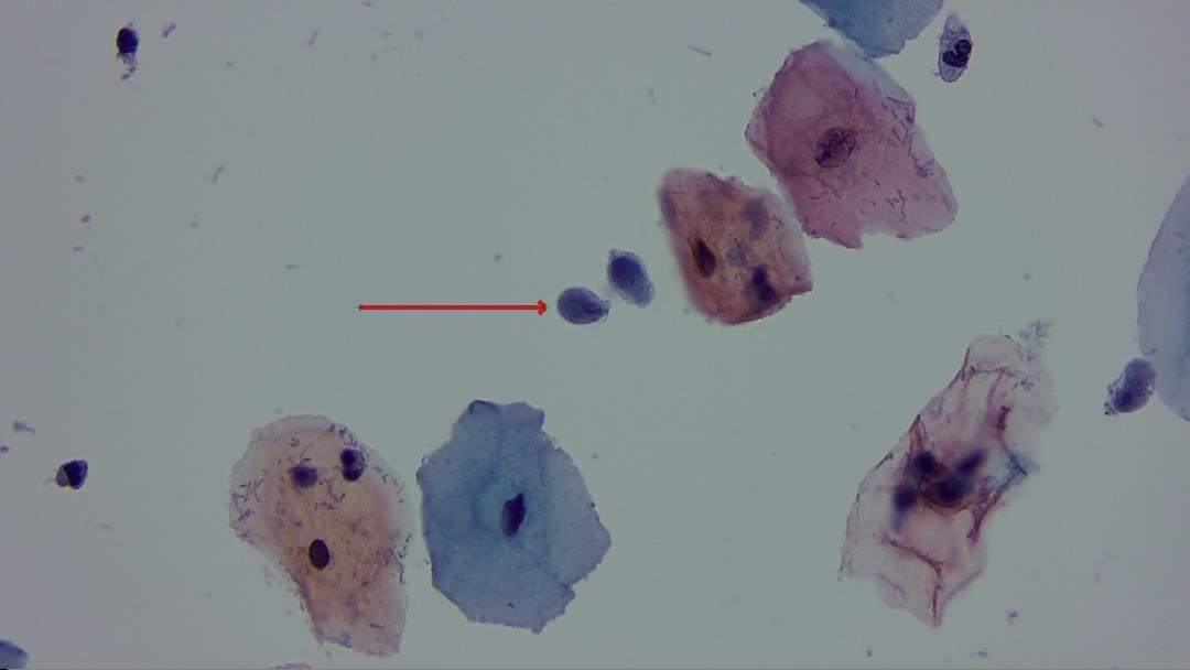
Fig. 6
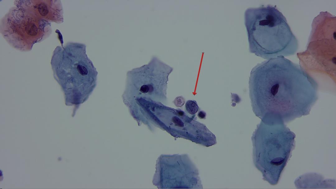
Fig. 7
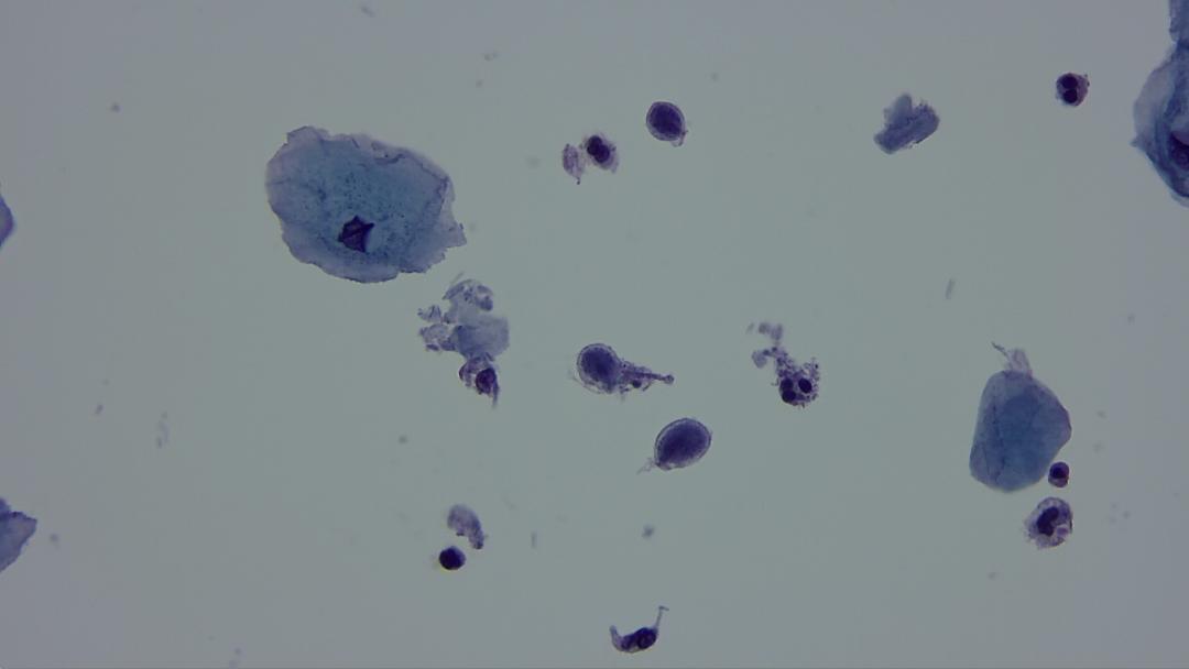
Fig. 8
It is imperative to recognize the subtle nuances of trichomonas morphology in liquid cytology samples. Their characteristic rounded form and absence of a typical dirty background, along with their frequent association with coccobacilli, set them apart from true cellular abnormalities. Failure to discern these features can lead to erroneous conclusions and subsequent patient mismanagement.
Incorporating advanced imaging technologies, such as the Motic Panthera, can aid in overcoming these diagnostic challenges. Its high-resolution capabilities and enhanced visualization empower clinicians to accurately identify and differentiate between true cellular abnormalities and benign mimics, such as trichomonas.
Ultimately, this case serves as a poignant reminder of the importance of meticulous observation and continued education in cytological interpretation. By honing our skills and embracing technological advancements, we can navigate the complexities of liquid-based cytology with confidence and precision, ensuring optimal patient care.
Copyright: Dr. Torres Gómez, Francisco Javier